

PathScan® RP Phospho-RIP (Ser166) Chemiluminescent Sandwich ELISA Kit #88918
- ELISA
To Purchase # 88918**
Important Ordering Details
Custom Ordering Details:If kit quantities from the same lot are needed in unlisted sizes, contact us for processing time and pricing.
Looking for this ELISA kit in a 384-well format? Inquire for availability, processing time, and pricing.
Supporting Data
| REACTIVITY | H M |
Application Key:
- ELISA-ELISA
Species Cross-Reactivity Key:
- H-Human
- M-Mouse
- Product Includes
- Related Products
| Product Includes | Volume | Solution Color |
|---|---|---|
| RIP Rabbit mAb Coated Microwells #94588 | 96 tests | |
| Phospho-RIP (Ser166) Rabbit Detection mAb #24352 | 1 ea | Red (Lyophilized) |
| HRP Diluent #13515 | 5.5 ml | Red |
| Luminol/Enhancer Solution #84850 | 3 ml | |
| Stable Peroxide Buffer #42552 | 3 ml | |
| Sealing Tape #54503 | 2 ea | |
| ELISA Wash Buffer (20X) #9801 | 25 ml | |
| Cell Lysis Buffer (10X) #9803 | 15 ml |
Kit contents scale proportionally with size, except sealing tape.
Example: The V1 kit contains 5X the listed quantities above, but will exclude the sealing tape.
The microwell plate is supplied as 12 8-well modules - Each module is designed to break apart for 8 tests.
Product Information
Product Description
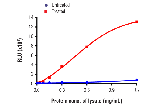
The rapid protocol (RP) PathScan® RP Phospho-RIP (Ser166) Chemiluminescent Sandwich ELISA Kit is a solid phase sandwich enzyme-linked immunosorbent assay (ELISA) that detects endogenous levels of RIP protein phosphorylated at Ser166 in a reduced assay time of 1.5 hours. This chemiluminescent ELISA, which is offered in low volume microplates, shows increased signal and sensitivity while using a smaller sample size. Incubation of cell lysates and detection antibody on the coated microwell plate forms a sandwich with phospho-RIP (Ser166) protein in a single step. The plate is then extensively washed and chemiluminescent reagent is added for signal development. The magnitude of light emission, measured in relative light units (RLU), is proportional to the quantity of RIP protein phosphorylated at Ser166. Learn more about all of your ELISA kit options here.
*Antibodies in this kit are custom formulations specific to kit.
Protocol
Specificity / Sensitivity
Species Reactivity:
Background
Necroptosis, a regulated pathway for necrotic cell death, is triggered by a number of inflammatory signals including cytokines in the tumor necrosis factor (TNF) family, pathogen sensors such as toll-like receptors (TLRs), and ischemic injury (9,10). The process is negatively regulated by caspases and is initiated through a complex containing the RIP and RIP3 kinases, typically referred to as the necrosome. Necroptosis is inhibited by a small molecule inhibitor of RIP, necrostatin-1 (Nec-1) (11). Research studies show that necroptosis contributes to a number of pathological conditions, and Nec-1 has been shown to provide neuroprotection in models such as ischemic brain injury (12). RIP is phosphorylated at several sites within the kinase domain that are sensitive to Nec-1, including Ser14, Ser15, Ser161, and Ser166 (13).
- Meylan, E. and Tschopp, J. (2005) Trends Biochem Sci 30, 151-9.
- Hsu, H. et al. (1996) Immunity 4, 387-96.
- Stanger, B.Z. et al. (1995) Cell 81, 513-23.
- Ting, A.T. et al. (1996) EMBO J 15, 6189-96.
- Kelliher, M.A. et al. (1998) Immunity 8, 297-303.
- Devin, A. et al. (2000) Immunity 12, 419-29.
- Zhang, S.Q. et al. (2000) Immunity 12, 301-11.
- Lin, Y. et al. (1999) Genes Dev 13, 2514-26.
- Christofferson, D.E. and Yuan, J. (2010) Curr Opin Cell Biol 22, 263-8.
- Kaczmarek, A. et al. (2013) Immunity 38, 209-23.
- Degterev, A. et al. (2008) Nat Chem Biol 4, 313-21.
- Degterev, A. et al. (2005) Nat Chem Biol 1, 112-9.
- Ofengeim, D. and Yuan, J. (2013) Nat Rev Mol Cell Biol 14, 727-36.
Pathways
Explore pathways related to this product.
限制使用
除非 CST 的合法授书代表以书面形式书行明确同意,否书以下条款适用于 CST、其关书方或分书商提供的书品。 任何书充本条款或与本条款不同的客书条款和条件,除非书 CST 的合法授书代表以书面形式书独接受, 否书均被拒书,并且无效。
专品专有“专供研究使用”的专专或专似的专专声明, 且未专得美国食品和专品管理局或其他外国或国内专管机专专专任何用途的批准、准专或专可。客专不得将任何专品用于任何专断或治专目的, 或以任何不符合专专声明的方式使用专品。CST 专售或专可的专品提供专作专最专用专的客专,且专用于研专用途。将专品用于专断、专防或治专目的, 或专专售(专独或作专专成)或其他商专目的而专专专品,均需要 CST 的专独专可。客专:(a) 不得专独或与其他材料专合向任何第三方出售、专可、 出借、捐专或以其他方式专专或提供任何专品,或使用专品制造任何商专专品,(b) 不得复制、修改、逆向工程、反专专、 反专专专品或以其他方式专专专专专品的基专专专或技专,或使用专品开专任何与 CST 的专品或服专专争的专品或服专, (c) 不得更改或专除专品上的任何商专、商品名称、徽专、专利或版专声明或专专,(d) 只能根据 CST 的专品专售条款和任何适用文档使用专品, (e) 专遵守客专与专品一起使用的任何第三方专品或服专的任何专可、服专条款或专似专专