

Phospho-Histone H3 (Ser28) Antibody #9713
Filter:
- WB
- IF
- F
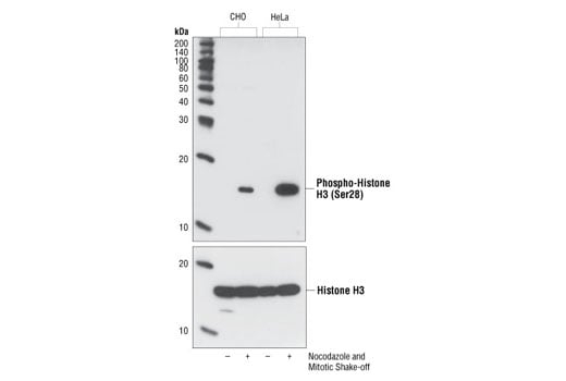
Western blot analysis of lysates from CHO and HeLa cells either untreated or synchronized in metaphase by treatment with 100 ng/ml nocodazole for 4 h, followed by isolation of metaphase cells by mitotic shake-off. Blots were probed with Phospho-Histone H3 (Ser28) Antibody #9713 (upper) or Histone H3 Antibody #9715 (lower).
Supporting Data
| REACTIVITY | H M Hm Dm |
| SENSITIVITY | Endogenous |
| MW (kDa) | 17 |
| SOURCE | Rabbit |
Application Key:
- WB-Western Blotting
- IF-Immunofluorescence
- F-Flow Cytometry
Species Cross-Reactivity Key:
- H-Human
- M-Mouse
- Hm-Hamster
- Dm-D. melanogaster
- Related Products
Product Information
Product Usage Information
| Application | Dilution |
|---|---|
| Western Blotting | 1:1000 |
| Immunofluorescence (Frozen) | 1:400 - 1:1600 |
| Immunofluorescence (Immunocytochemistry) | 1:400 - 1:1600 |
| Flow Cytometry (Fixed/Permeabilized) | 1:200 - 1:800 |
Storage
Supplied in 10 mM sodium HEPES (pH 7.5), 150 mM NaCl, 100 µg/ml BSA and 50% glycerol. Store at –20°C. Do not aliquot the antibody.
Protocol
Specificity / Sensitivity
Phospho-Histone H3 (Ser28) Antibody detects endogenous levels of histone H3 only when phosphorylated at Ser28. This antibody does not cross-react with other phosphorylated histones, including phospho-histone H3 (Ser10). Non-specific cytoplasmic staining may be observed by immunofluorescence in fixed-frozen tissues.
Species Reactivity:
Human, Mouse, Hamster, D. melanogaster
The antigen sequence used to produce this antibody shares 100% sequence homology with the species listed here, but reactivity has not been tested or confirmed to work by CST. Use of this product with these species is not covered under our Product Performance Guarantee.
Species predicted to react based on 100% sequence homology:
Rat, Chicken, Xenopus, Zebrafish, Bovine
Source / Purification
Polyclonal antibodies are produced by immunizing animals with a synthetic peptide corresponding to the amino terminus of histone H3 phosphorylated on Ser28. Antibodies are purified by protein A and peptide affinity chromatography.
Background
Modulation of chromatin structure plays an important role in the regulation of transcription in eukaryotes. The nucleosome, made up of DNA wound around eight core histone proteins (two each of H2A, H2B, H3, and H4), is the primary building block of chromatin (1). The amino-terminal tails of core histones undergo various posttranslational modifications, including acetylation, phosphorylation, methylation, and ubiquitination (2-5). These modifications occur in response to various stimuli and have a direct effect on the accessibility of chromatin to transcription factors and, therefore, gene expression (6). In most species, histone H2B is primarily acetylated at Lys5, 12, 15, and 20 (4,7). Histone H3 is primarily acetylated at Lys9, 14, 18, 23, 27, and 56. Acetylation of H3 at Lys9 appears to have a dominant role in histone deposition and chromatin assembly in some organisms (2,3). Phosphorylation at Ser10, Ser28, and Thr11 of histone H3 is tightly correlated with chromosome condensation during both mitosis and meiosis (8-10). Phosphorylation at Thr3 of histone H3 is highly conserved among many species and is catalyzed by the kinase haspin. Immunostaining with phospho-specific antibodies in mammalian cells reveals mitotic phosphorylation at Thr3 of H3 in prophase and its dephosphorylation during anaphase (11).
- Workman, J.L. and Kingston, R.E. (1998) Annu Rev Biochem 67, 545-79.
- Hansen, J.C. et al. (1998) Biochemistry 37, 17637-41.
- Strahl, B.D. and Allis, C.D. (2000) Nature 403, 41-5.
- Cheung, P. et al. (2000) Cell 103, 263-71.
- Bernstein, B.E. and Schreiber, S.L. (2002) Chem Biol 9, 1167-73.
- Jaskelioff, M. and Peterson, C.L. (2003) Nat Cell Biol 5, 395-9.
- Thorne, A.W. et al. (1990) Eur J Biochem 193, 701-13.
- Hendzel, M.J. et al. (1997) Chromosoma 106, 348-60.
- Goto, H. et al. (1999) J Biol Chem 274, 25543-9.
- Preuss, U. et al. (2003) Nucleic Acids Res 31, 878-85.
- Dai, J. et al. (2005) Genes Dev 19, 472-88.
Pathways
Explore pathways related to this product.
限制使用
除非 CST 的合法授书代表以书面形式书行明确同意,否书以下条款适用于 CST、其关书方或分书商提供的书品。 任何书充本条款或与本条款不同的客书条款和条件,除非书 CST 的合法授书代表以书面形式书独接受, 否书均被拒书,并且无效。
专品专有“专供研究使用”的专专或专似的专专声明, 且未专得美国食品和专品管理局或其他外国或国内专管机专专专任何用途的批准、准专或专可。客专不得将任何专品用于任何专断或治专目的, 或以任何不符合专专声明的方式使用专品。CST 专售或专可的专品提供专作专最专用专的客专,且专用于研专用途。将专品用于专断、专防或治专目的, 或专专售(专独或作专专成)或其他商专目的而专专专品,均需要 CST 的专独专可。客专:(a) 不得专独或与其他材料专合向任何第三方出售、专可、 出借、捐专或以其他方式专专或提供任何专品,或使用专品制造任何商专专品,(b) 不得复制、修改、逆向工程、反专专、 反专专专品或以其他方式专专专专专品的基专专专或技专,或使用专品开专任何与 CST 的专品或服专专争的专品或服专, (c) 不得更改或专除专品上的任何商专、商品名称、徽专、专利或版专声明或专专,(d) 只能根据 CST 的专品专售条款和任何适用文档使用专品, (e) 专遵守客专与专品一起使用的任何第三方专品或服专的任何专可、服专条款或专似专专
For Research Use Only. Not for Use in Diagnostic Procedures.
Cell Signaling Technology is a trademark of Cell Signaling Technology, Inc.
Alexa Fluor is a registered trademark of Life Technologies Corporation.
All other trademarks are the property of their respective owners. Visit our
Trademark Information page.