


Recombinant: Superior lot-to-lot consistency, continuous supply, and animal-free manufacturing.
Syndecan 1 (E7F7T) Rabbit mAb #96489
Filter:
- IHC
- F
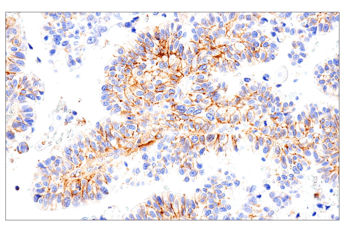
Immunohistochemical analysis of paraffin-embedded human urothelial carcinoma using Syndecan 1 (E7F7T) Rabbit mAb.
Supporting Data
| REACTIVITY | H |
| SENSITIVITY | Endogenous |
| MW (kDa) | |
| Source/Isotype | Rabbit IgG |
Application Key:
- IHC-Immunohistochemistry
- F-Flow Cytometry
Species Cross-Reactivity Key:
- H-Human
- Related Products
Product Information
Product Usage Information
| Application | Dilution |
|---|---|
| Immunohistochemistry (Paraffin) | 1:100 - 1:400 |
| Flow Cytometry (Fixed/Permeabilized) | 1:50 - 1:200 |
| Flow Cytometry (Live) | 1:50 - 1:200 |
Storage
Supplied in 10 mM sodium HEPES (pH 7.5), 150 mM NaCl, 100 µg/mL BSA, 50% glycerol, and less than 0.02% sodium azide. Store at –20°C. Do not aliquot the antibody.
For a carrier free (BSA and azide free) version of this product see product #25851.
For a carrier free (BSA and azide free) version of this product see product #25851.
Protocol
Specificity / Sensitivity
Syndecan 1 (E7F7T) Rabbit mAb recognizes endogenous levels of total syndecan 1 protein for approved applications. Non-specific staining was observed in skeletal muscle by immunohistochemistry.
Species Reactivity:
Human
Source / Purification
Monoclonal antibody is produced by immunizing animals with recombinant protein specific to the amino terminus in the extracellular region of human syndecan 1 protein.
Background
Syndecans are a family of type 1 transmembrane heparan sulfate proteoglycans comprising four members in mammals (SDC1-4) (1) encoded by four syndecan genes. Syndecans are involved in embryonic development, tumorigenesis, and angiogenesis (2). The extracellular domain harbors attachment sites for heparan sulfate and chondroitin sulfate chains, facilitating interaction with an array of proteins, including a plethora of growth factors. In addition, the hydrophobic C-terminal intracellular domain can interact with proteins containing a PDZ domain (2). These interactions place syndecans as important integrators of membrane signaling (3). Syndecans undergo proteolytic cleavage causing the release of their extracellular domain (shedding), converting the membrane-bound proteins into soluble molecular effectors (4).
Syndecan 1 (SDC1) is a specific marker for plasmacytic differentiation in hematologic disorders (5-7). This cell surface proteoglycan is also expressed in normal epithelial cells and tissues as well as various types of cancer tissues (8-11). The extracellular shed form of syndecan 1 remains soluble or accumulates in the extracellular matrix where it binds growth factors, cytokines and other extracellular matrix proteins (12,13). This binding activates signaling of bound growth factors or cytokines, which results in enhanced tumor growth, dissemination, angiogenesis, and osteolysis (14-17). As a result, the level of syndecan 1 protein and its shed form may serve as prognostic factors for a list of malignancies (6,18,19). Syndecan 1 has recently been found to be a critical mediator of macropinocytosis in pancreatic cancer (20).
Syndecan 1 (SDC1) is a specific marker for plasmacytic differentiation in hematologic disorders (5-7). This cell surface proteoglycan is also expressed in normal epithelial cells and tissues as well as various types of cancer tissues (8-11). The extracellular shed form of syndecan 1 remains soluble or accumulates in the extracellular matrix where it binds growth factors, cytokines and other extracellular matrix proteins (12,13). This binding activates signaling of bound growth factors or cytokines, which results in enhanced tumor growth, dissemination, angiogenesis, and osteolysis (14-17). As a result, the level of syndecan 1 protein and its shed form may serve as prognostic factors for a list of malignancies (6,18,19). Syndecan 1 has recently been found to be a critical mediator of macropinocytosis in pancreatic cancer (20).
- Couchman, J.R. (2003) Nat Rev Mol Cell Biol 4, 926-37.
- Multhaupt, H.A. et al. (2009) J Physiol Pharmacol 60 Suppl 4, 31-8.
- Zimmermann, P. and David, G. (1999) FASEB J 13 Suppl, S91-S100.
- Manon-Jensen, T. et al. (2010) FEBS J 277, 3876-89.
- Chilosi, M. et al. (1999) Mod Pathol 12, 1101-6.
- Seidel, C. et al. (2000) Blood 95, 388-92.
- O'Connell, F.P. et al. (2004) Am J Clin Pathol 121, 254-63.
- Inki, P. and Jalkanen, M. (1996) Ann Med 28, 63-7.
- Matsumoto, A. et al. (1997) Int J Cancer 74, 482-91.
- Conejo, J.R. et al. (2000) Int J Cancer 88, 12-20.
- Zellweger, T. et al. (2003) Prostate 55, 20-9.
- Bayer-Garner, I.B. et al. (2001) Mod Pathol 14, 1052-8.
- Ramani, V.C. et al. (2013) FEBS J 280, 2294-306.
- Derksen, P.W. et al. (2002) Blood 99, 1405-10.
- You, W.K. and McDonald, D.M. (2008) BMB Rep 41, 833-9.
- Ramani, V.C. et al. (2011) J Biol Chem 286, 6490-9.
- Aragão, A.Z. et al. (2012) PLoS One 7, e43521.
- Joensuu, H. et al. (2002) Cancer Res 62, 5210-7.
- Lee, S.H. et al. (2014) Int J Clin Oncol 19, 247-53.
- Yao, W. et al. (2019) Nature 568, 410-414.
限制使用
除非 CST 的合法授书代表以书面形式书行明确同意,否书以下条款适用于 CST、其关书方或分书商提供的书品。 任何书充本条款或与本条款不同的客书条款和条件,除非书 CST 的合法授书代表以书面形式书独接受, 否书均被拒书,并且无效。
专品专有“专供研究使用”的专专或专似的专专声明, 且未专得美国食品和专品管理局或其他外国或国内专管机专专专任何用途的批准、准专或专可。客专不得将任何专品用于任何专断或治专目的, 或以任何不符合专专声明的方式使用专品。CST 专售或专可的专品提供专作专最专用专的客专,且专用于研专用途。将专品用于专断、专防或治专目的, 或专专售(专独或作专专成)或其他商专目的而专专专品,均需要 CST 的专独专可。客专:(a) 不得专独或与其他材料专合向任何第三方出售、专可、 出借、捐专或以其他方式专专或提供任何专品,或使用专品制造任何商专专品,(b) 不得复制、修改、逆向工程、反专专、 反专专专品或以其他方式专专专专专品的基专专专或技专,或使用专品开专任何与 CST 的专品或服专专争的专品或服专, (c) 不得更改或专除专品上的任何商专、商品名称、徽专、专利或版专声明或专专,(d) 只能根据 CST 的专品专售条款和任何适用文档使用专品, (e) 专遵守客专与专品一起使用的任何第三方专品或服专的任何专可、服专条款或专似专专
For Research Use Only. Not For Use In Diagnostic Procedures.
Cell Signaling Technology is a trademark of Cell Signaling Technology, Inc.
All other trademarks are the property of their respective owners. Visit our
Trademark Information page.