

TGF-β Fibrosis Pathway Antibody Sampler Kit #77397
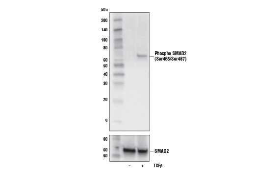
Western blot analysis of extracts from HaCaT cells, untreated (-) or treated with Human Transforming Growth Factor β3 (hTGF-β3) #8425 (100ng/ml, 30mins) (+), using Phospho SMAD2 (Ser465/Ser467) (E8F3R) Rabbit mAb (upper) and total Smad2 (D43B4) XP® Rabbit mAb, #5339 (lower).
- Product Includes
- Related Products
| Product Includes | Quantity | Applications | Reactivity | MW(kDa) | Isotype |
|---|---|---|---|---|---|
| α-Smooth Muscle Actin (D4K9N) XP® Rabbit mAb #19245 | 20 µl | WB, IP, IHC, IF | H M R Hm Mk | 42 | Rabbit IgG |
| COL1A1 (E8I9Z) Rabbit mAb #91144 | 20 µl | WB, IP | H | 220 | Rabbit IgG |
| SMAD2/3 (D7G7) XP® Rabbit mAb #8685 | 20 µl | WB, IP, IF, F, ChIP | H M R Mk | 52, 60 | Rabbit IgG |
| SMAD2 (D43B4) XP® Rabbit mAb #5339 | 20 µl | WB, IP, IF, F, ChIP | H M R Mk | 60 | Rabbit IgG |
| Phospho-SMAD2 (Ser465/Ser467) (E8F3R) Rabbit mAb #18338 | 20 µl | WB, IP, IF, F, ChIP | H M R | 60 | Rabbit IgG |
| YKL-40 (E2L1M) Rabbit mAb #47066 | 20 µl | WB, IHC, IF | H | 30-40 | Rabbit IgG |
| Phospho-SMAD2 (Ser465/467)/SMAD3 (Ser423/425) (D27F4) Rabbit mAb #8828 | 20 µl | WB | H M R Mk | 52, 60 | Rabbit IgG |
| TGF-β (56E4) Rabbit mAb #3709 | 20 µl | WB | H | 12, 45-60 | Rabbit IgG |
| TGF-β Receptor II (E5M6F) Rabbit mAb #41896 | 20 µl | WB | H | 85 | Rabbit IgG |
| Anti-rabbit IgG, HRP-linked Antibody #7074 | 100 µl | WB | Rab | Goat |
Product Information
Kit Usage Information
Protocols
- 3709: Western Blotting
- 5339: Western Blotting, Immunoprecipitation (Agarose), Immunofluorescence, Flow, ChIP Magnetic
- 7074: Western Blotting
- 8685: Western Blotting, Immunoprecipitation (Agarose), Immunofluorescence, Flow, ChIP Magnetic, Chromatin IP-seq
- 8828: Western Blotting
- 18338: Western Blotting, Fluorescent Western, Immunoprecipitation (Agarose), Immunofluorescence, Flow, ChIP Magnetic
- 19245: Western Blotting, Immunoprecipitation (Magnetic), Immunohistochemistry (Leica® Bond™), Immunohistochemistry (Paraffin), Immunofluorescence
- 41896: Western Blotting
- 47066: Western Blotting, Immunohistochemistry (Paraffin), Immunofluorescence
- 91144: Western Blotting, Immunoprecipitation (Magnetic)
Product Description
The TGF-β Fibrosis Pathway Antibody Sampler Kit provides an economical means of investigating activation of TGF-β/ SMAD2/3 signaling pathways in cells or tissues that lead to the expression of profibrotic genes, including expression of α-Smooth Muscle Actin in activated fibroblasts, and upregulation of Collagen1A1, Col11A1, and YKL-40. The kit includes enough antibodies to perform at least two western blot experiments with each primary antibody.
Background
Transforming growth factor-β (TGF-β) superfamily members are critical regulators of cell proliferation and differentiation, developmental patterning and morphogenesis, and disease pathogenesis (1-4). In the context of fibrosis, TGF-β signaling to SMAD2/3 is one of the biggest drivers of the profibrotic program (5).
TGF-β elicits signaling through three cell surface receptors: type I (RI), type II (RII), and type III (RIII). In response to ligand binding, the type II receptors form stable heterotrimeric complexes with the type I receptors, allowing phosphorylation and activation of type I receptor kinase. Activated type I receptors associate with SMAD2/3 and phosphorylate them on a conserved carboxy terminal SSXS motif. The phosphorylated SMADs dissociate from the receptor and form a heterotrimeric complex with the co-Smad (Smad4), allowing translocation of the complex to the nucleus. Once in the nucleus, phosphorylated SMAD2/3 targets a subset of DNA binding proteins to regulate the transcriptional program (6-8).
In the context of fibrosis, SMAD2/3 activation upregulates expression of profibrotic genes such as COL1A1 and other ECM modulators that modify the extracellular matrix of the tissue. (9). TGF-β/ SMAD2/3 signaling also induces expression of α-Smooth Muscle Actin in fibroblasts, causing transformation of these cells to myofibroblasts (10). Myofibroblasts further modify the ECM, causing excessive accumulation of collagens and other ECM components. Injury to the tissue attracts macrophages and other immune cells and the fibrotic tissue soon becomes a site of inflammation (11). In this pro-fibrotic, pro-inflammatory environment, YKL-40, also known as Chitinase-3-like protein 1 (CHI3L1), is secreted. YKL-40 is a pro-inflammatory glycoprotein that also contributes to the progression of fibrosis (12). Measurement of collagen content, α-Smooth Muscle Actin, and the release of YKL-40 are predictive of fibrotic activity.
- Massagué, J. et al. (2000) Cell 103, 295-309.
- de Caestecker, M.P. et al. (2000) J Natl Cancer Inst 92, 1388-402.
- Derynck, R. et al. (2001) Nat Genet 29, 117-29.
- Miyazono, K. et al. (2000) Adv Immunol 75, 115-57.
- Meng, X.M. et al. (2016) Nat Rev Nephrol 12, 325-38.
- Wu, G. et al. (2000) Science 287, 92-7.
- Attisano, L. and Wrana, J.L. (2002) Science 296, 1646-7.
- Moustakas, A. et al. (2001) J Cell Sci 114, 4359-69.
- Bagalad, B.S. et al. J Oral Maxillofac Pathol 21, 462-3.
- Mack, M. (2018) Matrix Biol 68-69, 106-21.
- Johansen, J.S. (2006) Dan Med Bull 53, 172-209.
Pathways
Explore pathways related to this product.
限制使用
除非 CST 的合法授书代表以书面形式书行明确同意,否书以下条款适用于 CST、其关书方或分书商提供的书品。 任何书充本条款或与本条款不同的客书条款和条件,除非书 CST 的合法授书代表以书面形式书独接受, 否书均被拒书,并且无效。
专品专有“专供研究使用”的专专或专似的专专声明, 且未专得美国食品和专品管理局或其他外国或国内专管机专专专任何用途的批准、准专或专可。客专不得将任何专品用于任何专断或治专目的, 或以任何不符合专专声明的方式使用专品。CST 专售或专可的专品提供专作专最专用专的客专,且专用于研专用途。将专品用于专断、专防或治专目的, 或专专售(专独或作专专成)或其他商专目的而专专专品,均需要 CST 的专独专可。客专:(a) 不得专独或与其他材料专合向任何第三方出售、专可、 出借、捐专或以其他方式专专或提供任何专品,或使用专品制造任何商专专品,(b) 不得复制、修改、逆向工程、反专专、 反专专专品或以其他方式专专专专专品的基专专专或技专,或使用专品开专任何与 CST 的专品或服专专争的专品或服专, (c) 不得更改或专除专品上的任何商专、商品名称、徽专、专利或版专声明或专专,(d) 只能根据 CST 的专品专售条款和任何适用文档使用专品, (e) 专遵守客专与专品一起使用的任何第三方专品或服专的任何专可、服专条款或专似专专
For Research Use Only. Not for Use in Diagnostic Procedures.
Cell Signaling Technology is a trademark of Cell Signaling Technology, Inc.
CST is a registered trademark of Cell Signaling Technology, Inc.
XP is a registered trademark of Cell Signaling Technology, Inc.
U.S. Patent No. 5,675,063.
All other trademarks are the property of their respective owners. Visit our
Trademark Information page.