


Recombinant: Superior lot-to-lot consistency, continuous supply, and animal-free manufacturing.
TNF-R1 (C25C1) Rabbit mAb #3736
Filter:
- WB
- IP
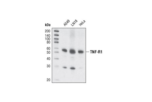
Western blot analysis of extracts from A549, LN-18 and HeLa cells using TNF-R1 (C25C1) Rabbit mAb.
Supporting Data
| REACTIVITY | H |
| SENSITIVITY | Endogenous |
| MW (kDa) | 55 |
| Source/Isotype | Rabbit IgG |
Application Key:
- WB-Western Blotting
- IP-Immunoprecipitation
Species Cross-Reactivity Key:
- H-Human
- Related Products
Product Information
Product Usage Information
| Application | Dilution |
|---|---|
| Western Blotting | 1:1000 |
| Simple Western™ | 1:10 - 1:50 |
| Immunoprecipitation | 1:100 |
Storage
Supplied in 10 mM sodium HEPES (pH 7.5), 150 mM NaCl, 100 µg/ml BSA, 50% glycerol and less than 0.02% sodium azide. Store at –20°C. Do not aliquot the antibody.
Protocol
Specificity / Sensitivity
TNF-R1 (C25C1) Rabbit mAb recognizes endogenous levels of human TNF-R1 protein. This antibody does not appear to cross-react with other related family members. This antibody may recognize a 30 kDa splice isoform of TNF-R1 in some cell lines.
Species Reactivity:
Human
Source / Purification
Monoclonal antibody is produced by immunizing animals with a synthetic peptide corresponding to residues within the intracellular region and surrounding Ser331 of human TNF-R1 protein.
Background
TNF-α is an important cytokine produced by numerous cell types, including neutrophils, activated lymphocytes, macrophages, and NK cells. It plays a critical role in inflammatory responses and apoptosis (1). TNF-α exists as a membrane-anchored and soluble form, both of which show biological activity. Response to TNF-α is mediated through two receptors, TNF-R1, which is widely expressed, and TNF-R2, which is expressed mainly in immune and endothelial cells (2). Antagonists to TNF-α have been validated as therapeutic targets for rheumatoid arthritis and other immune disorders (3).
The two receptors for TNF-α, TNF-R1 (55 kDa) and TNF-R2 (75 kDa) can mediate distinct cellular responses (4,5). In most cases cytotoxicity elicited by TNF has been reported to act through TNF-R1 (6,7). Cytotoxicity is mediated by a "death domain" with the intracellular region of the receptor that binds to the death domain adaptor protein TRADD and triggers the activation of caspases (8). Soluble forms of both receptors have also been characterized which can bind TNF-α and may play an important role in immune disorders (9,10).
The two receptors for TNF-α, TNF-R1 (55 kDa) and TNF-R2 (75 kDa) can mediate distinct cellular responses (4,5). In most cases cytotoxicity elicited by TNF has been reported to act through TNF-R1 (6,7). Cytotoxicity is mediated by a "death domain" with the intracellular region of the receptor that binds to the death domain adaptor protein TRADD and triggers the activation of caspases (8). Soluble forms of both receptors have also been characterized which can bind TNF-α and may play an important role in immune disorders (9,10).
- Aggarwal, B.B. (2003) Nat Rev Immunol 3, 745-56.
- Locksley, R.M. et al. (2001) Cell 104, 487-501.
- Taylor, P.C. et al. (2004) Curr Opin Biotechnol 15, 557-63.
- Tartaglia, L.A. et al. (1991) Proc Natl Acad Sci USA 88, 9292-6.
- Peschon, J.J. et al. (1998) J Immunol 160, 943-52.
- Tartaglia, L.A. et al. (1993) Cell 73, 213-6.
- Rothe, J. et al. (1993) Nature 364, 798-802.
- Chen, G. and Goeddel, D.V. (2002) Science 296, 1634-5.
- Humbert, M. et al. (1994) Am J Respir Crit Care Med 149, 1681-5.
- Schröder, J. et al. Infection 23, 143-8.
Pathways
Explore pathways related to this product.
限制使用
除非 CST 的合法授书代表以书面形式书行明确同意,否书以下条款适用于 CST、其关书方或分书商提供的书品。 任何书充本条款或与本条款不同的客书条款和条件,除非书 CST 的合法授书代表以书面形式书独接受, 否书均被拒书,并且无效。
专品专有“专供研究使用”的专专或专似的专专声明, 且未专得美国食品和专品管理局或其他外国或国内专管机专专专任何用途的批准、准专或专可。客专不得将任何专品用于任何专断或治专目的, 或以任何不符合专专声明的方式使用专品。CST 专售或专可的专品提供专作专最专用专的客专,且专用于研专用途。将专品用于专断、专防或治专目的, 或专专售(专独或作专专成)或其他商专目的而专专专品,均需要 CST 的专独专可。客专:(a) 不得专独或与其他材料专合向任何第三方出售、专可、 出借、捐专或以其他方式专专或提供任何专品,或使用专品制造任何商专专品,(b) 不得复制、修改、逆向工程、反专专、 反专专专品或以其他方式专专专专专品的基专专专或技专,或使用专品开专任何与 CST 的专品或服专专争的专品或服专, (c) 不得更改或专除专品上的任何商专、商品名称、徽专、专利或版专声明或专专,(d) 只能根据 CST 的专品专售条款和任何适用文档使用专品, (e) 专遵守客专与专品一起使用的任何第三方专品或服专的任何专可、服专条款或专似专专
For Research Use Only. Not For Use In Diagnostic Procedures.
Cell Signaling Technology is a trademark of Cell Signaling Technology, Inc.
U.S. Patent No. 7,429,487, foreign equivalents, and child patents deriving therefrom.
All other trademarks are the property of their respective owners. Visit our
Trademark Information page.