


Recombinant: Superior lot-to-lot consistency, continuous supply, and animal-free manufacturing.
VISTA (D1L2G™) XP® Rabbit mAb #64953
Filter:
- WB
- IHC
- F
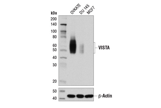
Western blot analysis of extracts from OVKATE, DU 145, and MCF7 cells using VISTA ((D1L2G™) XP® Rabbit mAb (upper) or β-Actin (D6A8) Rabbit mAb #8457 (lower).
Supporting Data
| REACTIVITY | H Mk |
| SENSITIVITY | Endogenous |
| MW (kDa) | 45-65 |
| Source/Isotype | Rabbit IgG |
Application Key:
- WB-Western Blotting
- IHC-Immunohistochemistry
- F-Flow Cytometry
Species Cross-Reactivity Key:
- H-Human
- Mk-Monkey
- Related Products
- Conjugates
Product Information
Product Usage Information
| Application | Dilution |
|---|---|
| Western Blotting | 1:1000 |
| IHC Leica Bond | 1:100 - 1:400 |
| Immunohistochemistry (Paraffin) | 1:100 - 1:400 |
| Flow Cytometry (Fixed/Permeabilized) | 1:50 - 1:200 |
Storage
Supplied in 10 mM sodium HEPES (pH 7.5), 150 mM NaCl, 100 µg/ml BSA, 50% glycerol and less than 0.02% sodium azide. Store at –20°C. Do not aliquot the antibody.
For a carrier-free (BSA and azide free) version of this product see product #82119.
For a carrier-free (BSA and azide free) version of this product see product #82119.
Protocol
Specificity / Sensitivity
VISTA (D1L2G™) XP® Rabbit mAb recognizes endogenous levels of total VISTA protein. Non-specific staining was observed in kidney and prostate epithelium by immunohistochemistry.
Species Reactivity:
Human, Monkey
Source / Purification
Monoclonal antibody is produced by immunizing animals with a synthetic peptide corresponding to residues near the carboxy terminus of human VISTA protein.
Background
VISTA (V-Domain Ig Suppressor of T Cell Activation) is a negative checkpoint control protein that regulates T cell activation and immune responses. VISTA, which contains a single Ig-like V-type domain, a transmembrane domain, and an intracellular domain, has sequence similarity to both the B7 and CD28 family members. Although primarily expressed by myeloid cells, VISTA is also expressed by CD4+, CD8+, and FoxP3+ T-cells. Thus, VISTA is described as both a ligand and a receptor (1-3). Blocking VISTA induces T-cell activation and proliferation, and potentiates disease severity in the EAE model (1). Furthermore, genetic deletion of VISTA in mice leads to spontaneous T-cell activation and chronic inflammation (4,5). In mouse models of cancer, neutralization of VISTA enhances T-cell proliferation and effector function and increases tumor infiltration, suggesting VISTA blockade could be an effective strategy for tumor immunotherapy (6,7).
- Wang, L. et al. (2011) J Exp Med 208, 577-92.
- Flies, D.B. et al. (2011) J Immunol 187, 1537-41.
- Lines, J.L. et al. (2014) Cancer Res 74, 1924-32.
- Wang, L. et al. (2014) Proc Natl Acad Sci U S A 111, 14846-51.
- Liu, J. et al. (2015) Proc Natl Acad Sci U S A 112, 6682-7.
- Le Mercier, I. et al. (2014) Cancer Res 74, 1933-44.
- Lines, J.L. et al. (2014) Cancer Immunol Res 2, 510-7.
限制使用
除非 CST 的合法授书代表以书面形式书行明确同意,否书以下条款适用于 CST、其关书方或分书商提供的书品。 任何书充本条款或与本条款不同的客书条款和条件,除非书 CST 的合法授书代表以书面形式书独接受, 否书均被拒书,并且无效。
专品专有“专供研究使用”的专专或专似的专专声明, 且未专得美国食品和专品管理局或其他外国或国内专管机专专专任何用途的批准、准专或专可。客专不得将任何专品用于任何专断或治专目的, 或以任何不符合专专声明的方式使用专品。CST 专售或专可的专品提供专作专最专用专的客专,且专用于研专用途。将专品用于专断、专防或治专目的, 或专专售(专独或作专专成)或其他商专目的而专专专品,均需要 CST 的专独专可。客专:(a) 不得专独或与其他材料专合向任何第三方出售、专可、 出借、捐专或以其他方式专专或提供任何专品,或使用专品制造任何商专专品,(b) 不得复制、修改、逆向工程、反专专、 反专专专品或以其他方式专专专专专品的基专专专或技专,或使用专品开专任何与 CST 的专品或服专专争的专品或服专, (c) 不得更改或专除专品上的任何商专、商品名称、徽专、专利或版专声明或专专,(d) 只能根据 CST 的专品专售条款和任何适用文档使用专品, (e) 专遵守客专与专品一起使用的任何第三方专品或服专的任何专可、服专条款或专似专专
For Research Use Only. Not For Use In Diagnostic Procedures.
Cell Signaling Technology is a trademark of Cell Signaling Technology, Inc.
XP is a registered trademark of Cell Signaling Technology, Inc.
All other trademarks are the property of their respective owners. Visit our
Trademark Information page.